Abstract
Background: Ibogaine was recently found to result in significant functional improvements in treating the sequelae of traumatic brain injury (TBI) among Special Operations Forces veterans (SOVs). In the present article, we use multimodal neuroimaging to elucidate the neural correlates of ibogaine in 30 male SOVs who received ibogaine treatment.
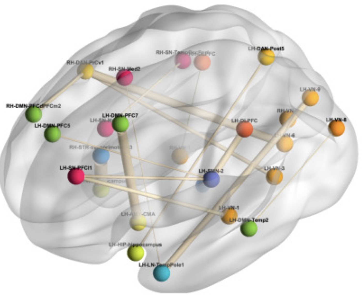
Methods: Arterial spin labeling and blood oxygen level-dependent functional magnetic resonance imaging data were collected before, immediately after ibogaine treatment, and at 1-month follow-up. A whole-brain exploratory analysis was conducted to examine the effects of ibogaine on resting-state regional cerebral blood flow (rCBF) and functional connectivity.
Results: The results revealed gradual increases in rCBF in the cortical, limbic, and striatal subregions, and changes in functional connectivity across a wide range of functional networks. The magnitude of treatment-induced rCBF changes in the left insula and left anterior cingulate cortex correlated significantly with improvements in TBI-related disability symptoms.
Conclusion: Our results suggest that ibogaine may involve widespread reorganization of functional connections in the brain, and that persisting regional changes in metabolic activity after ibogaine treatment, particularly within paralimbic brain regions, might be related to the observed therapeutic effects of ibogaine. Our findings serve to generate future hypotheses for larger, controlled neuroimaging studies of ibogaine in humans, necessary to validate these initial findings.