Abstract
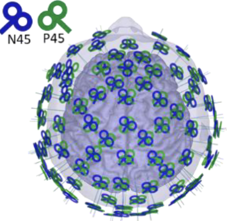
Large-scale networks underpin brain functions. How such networks respond to focal stimulation can help decipher complex brain processes and optimize brain stimulation treatments. To map such stimulation-response patterns across the brain non-invasively, we recorded concurrent EEG responses from single-pulse transcranial magnetic stimulation (i.e., TMS-EEG) from over 100 cortical regions with two orthogonal coil orientations from one densely-sampled individual. We also acquired Human Connectome Project (HCP)-styled diffusion imaging scans (six), resting- state functional Magnetic Resonance Imaging (fMRI) scans (120 mins), resting-state EEG scans (108 mins), and structural MR scans (T1- and T2-weighted). Using the TMS-EEG data, we applied network science-based community detection to reveal insights about the brain’s causal-functional organization from both a stimulation and recording perspective. We also computed structural and functional maps and the electric field of each TMS stimulation condition. Altogether, we hope the release of this densely sampled (n=1) dataset will be a uniquely valuable resource for both basic and clinical neuroscience research.